
Specjalizacje
-
Okulistyka
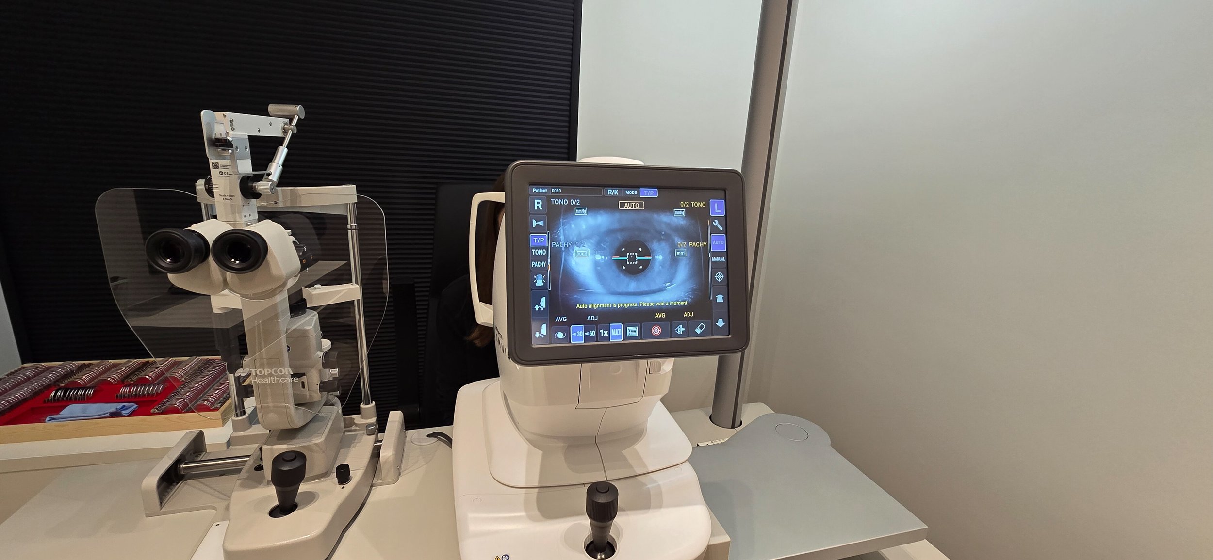
W ramach poradni okulistycznej oferujemy kompleksową diagnostykę narządu wzroku, opartą na nowoczesnym sprzęcie oraz doświadczeniu naszych specjalistów. Wykonujemy szereg badań pozwalających na dokładną ocenę stanu oka oraz wczesne wykrycie wielu schorzeń.
-
Elektrofizjologia
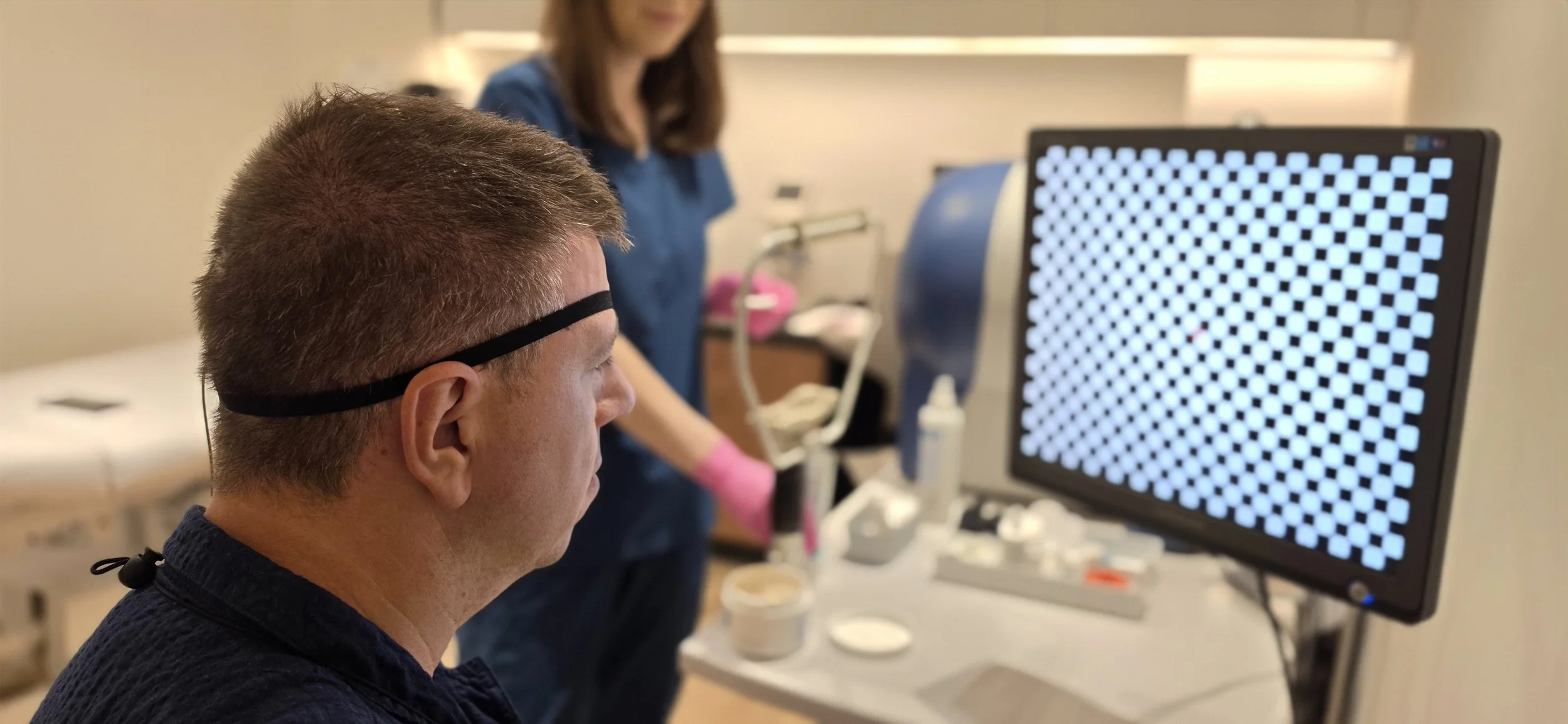
Badania elektrofizjologiczne narządu wzroku to specjalistyczne badania czynnościowe, które umożliwiają ocenę funkcjonowania siatkówki oraz nerwu wzrokowego. Stanowią ważny element diagnostyki okulistycznej.
-
USG Doppler
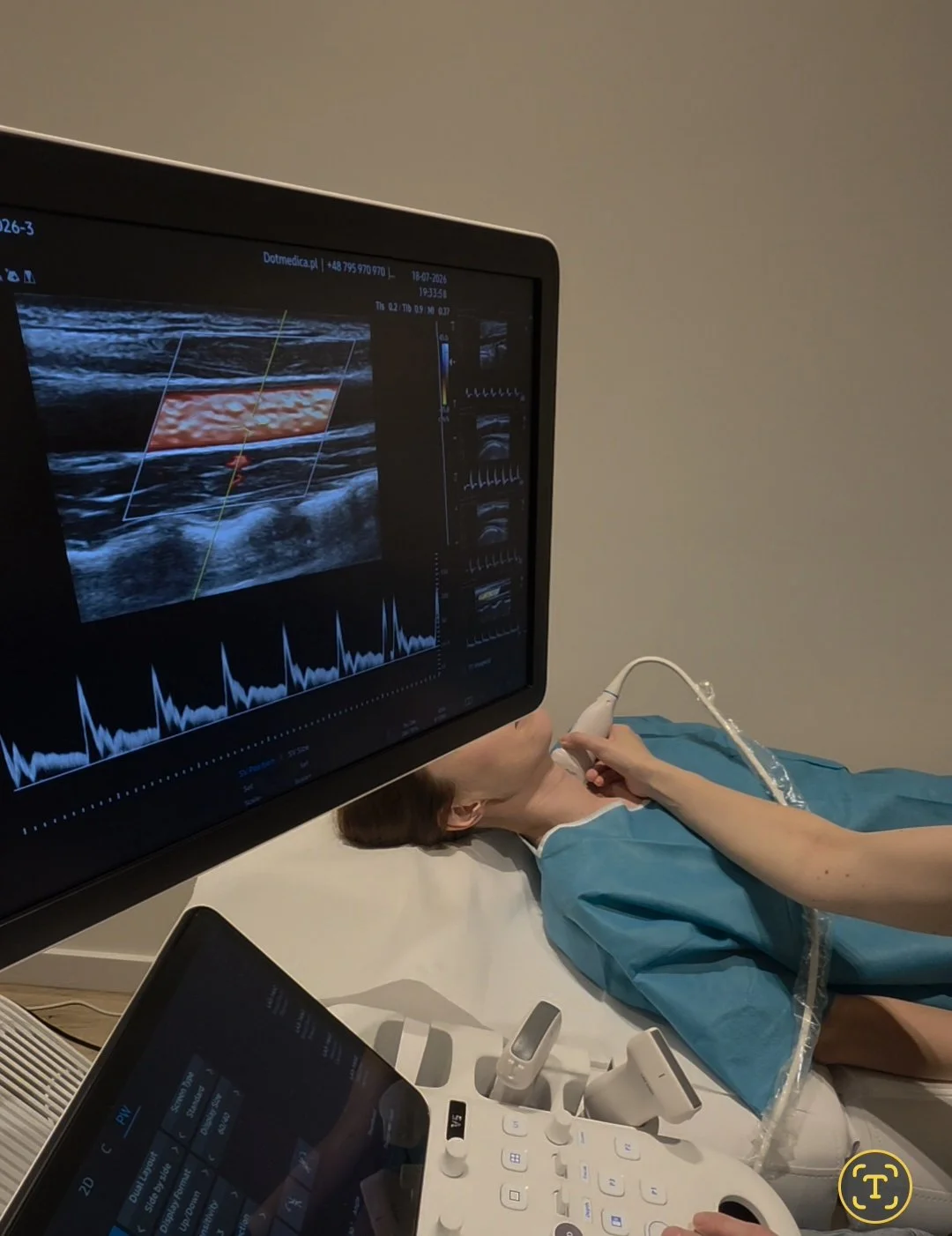
USG Doppler to nieinwazyjne i bezpieczne badanie ultrasonograficzne, które pozwala ocenić przepływ krwi w tętnicach i żyłach. Dzięki zastosowaniu efektu Dopplera możliwe jest wykrycie zaburzeń krążenia oraz ocena drożności i wydolności naczyń krwionośnych.
Okulistyka - OCT
Badanie OCT
-
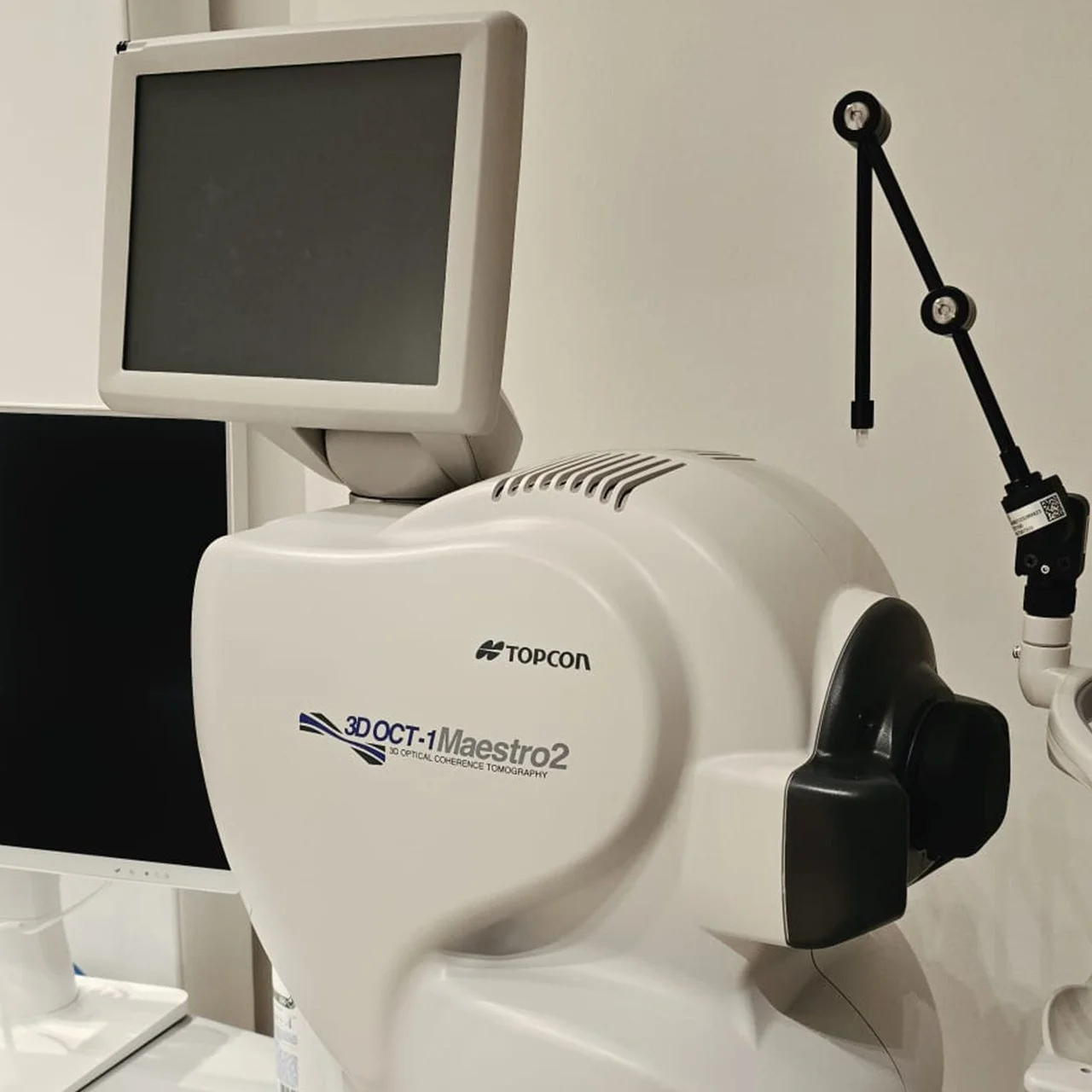
OCT to nowoczesne, nieinwazyjne badanie obrazowe pozwalające na bardzo dokładną ocenę struktur oka – przede wszystkim siatkówki (w tym plamki żółtej), nerwu wzrokowego oraz rogówki.
Badanie jest szybkie, nie wymaga kontaktu urządzenia z okiem i zapewnia bardzo precyzyjne obrazy jego struktur.
Przygotowanie: badanie zwykle trwa około 10 min
Przed badaniem najczęściej rozszerza się źrenicę badanego oka.
-
Badanie OCT plamki lub nerwu (obuocznie) - 260,00 zł
Badanie OCT plamki i nerwu (obuocznie) - 300,00 zł
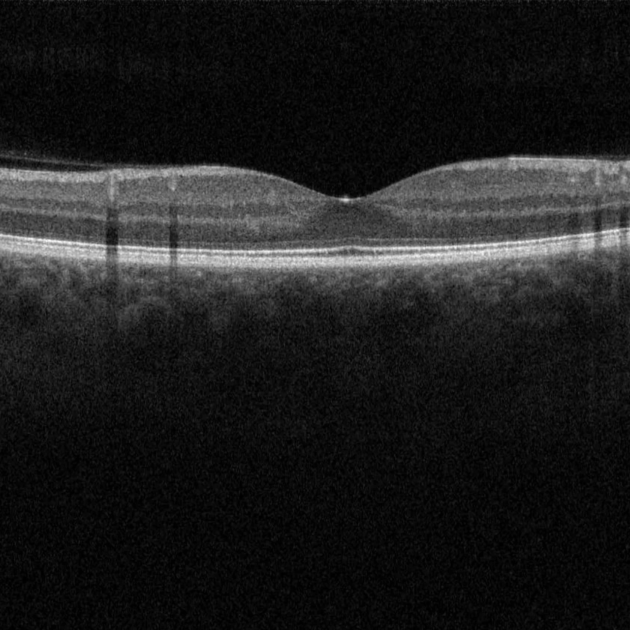
Badanie OCT plamki
-
OCT plamki, czyli tomografia optyczna plamki żółtej, to niezwykle cenne badanie w diagnostyce chorób siatkówki, szczególnie w obrębie plamki.
OCT jest szczególnie przydatne w diagnostyce chorób plamki, takich jak zwyrodnienie plamki związane z wiekiem - AMD, a także w rozpoznawaniu obrzęku plamki wynikającego miedzy innymi z cukrzycy lub innych schorzeń.
-
Badanie OCT plamki (obuocznie) - 260,00 zł
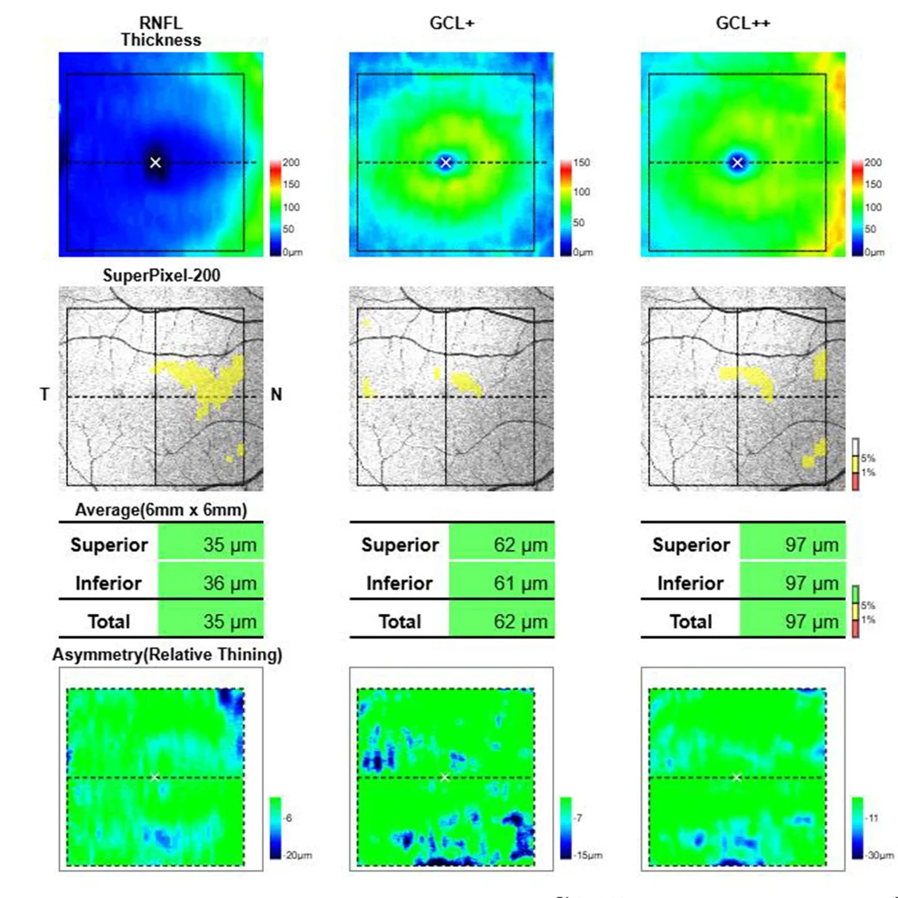
Badanie OCT nerwu / RNFL / GCC
-
RNFL oraz GCC, często określane też jako OCT nerwu wzrokowego lub wykonywane celem wykrycia jaskry.
OCT RNFL umożliwia analizę grubości warstwy włókien nerwowych, co ma kluczowe znaczenie w diagnostyce jaskry oraz innych chorób wpływających na nerw wzrokowy. Z kolei badanie GCC pozwala ocenić warstwę komórek zwojowych, co umożliwia bardzo wczesne wykrywanie ich uszkodzeń.
-
Badanie OCT nerwu (obuocznie) - 260,00 zł
Okulistyka - Badania
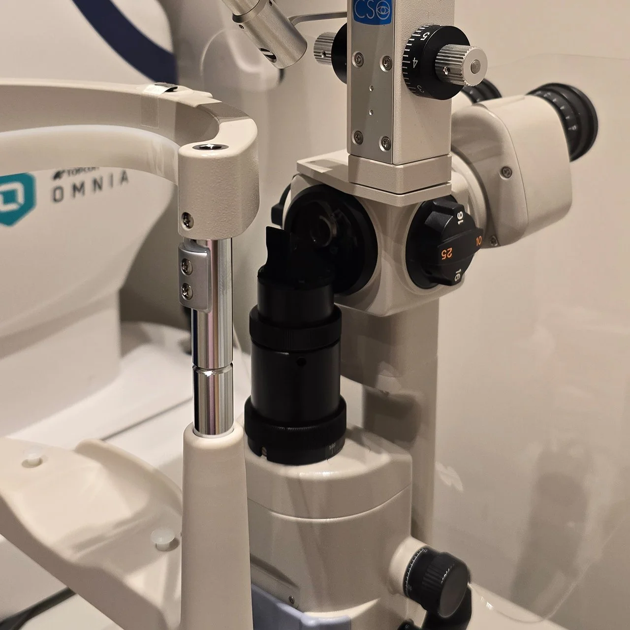
Badanie w lampie szczelinowej
-
Podstawowe badanie okulistyczne umożliwiające szczegółową ocenę przedniego odcinka oka, a przy użyciu dodatkowych soczewek także tylnego odcinka, w tym dna oka.
Stanowi standardowy element każdej wizyty okulistycznej.
Badanie dna oka wykonywane jest po podaniu kropli rozszerzających źrenice.
-
Badanie jest wliczone w koszt konsultacji okulistycznej
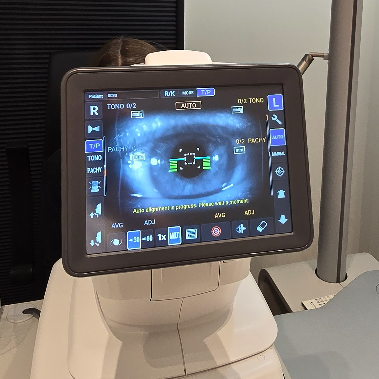
Autorefraktometria
-
Autorefraktometria to szybkie, bezbolesne i w pełni zautomatyzowane badanie, które pozwala wstępnie ocenić wadę wzroku. W trakcie badania wystarczy spojrzeć na wskazany obraz w. Uzyskany wynik stanowi punkt wyjścia do dalszego, dokładniejszego badania wzroku oraz precyzyjnego doboru okularów lub soczewek kontaktowych.
W naszym gabinecie korzystamy z nowoczesnego urządzenia, które podczas jednego badania umożliwia również ocenę krzywizny rogówki (keratometria), pomiar ciśnienia wewnątrzgałkowego oraz grubości rogówki.
-
Badanie jest wliczone w koszt konsultacji okulistycznej
Tonometria
-
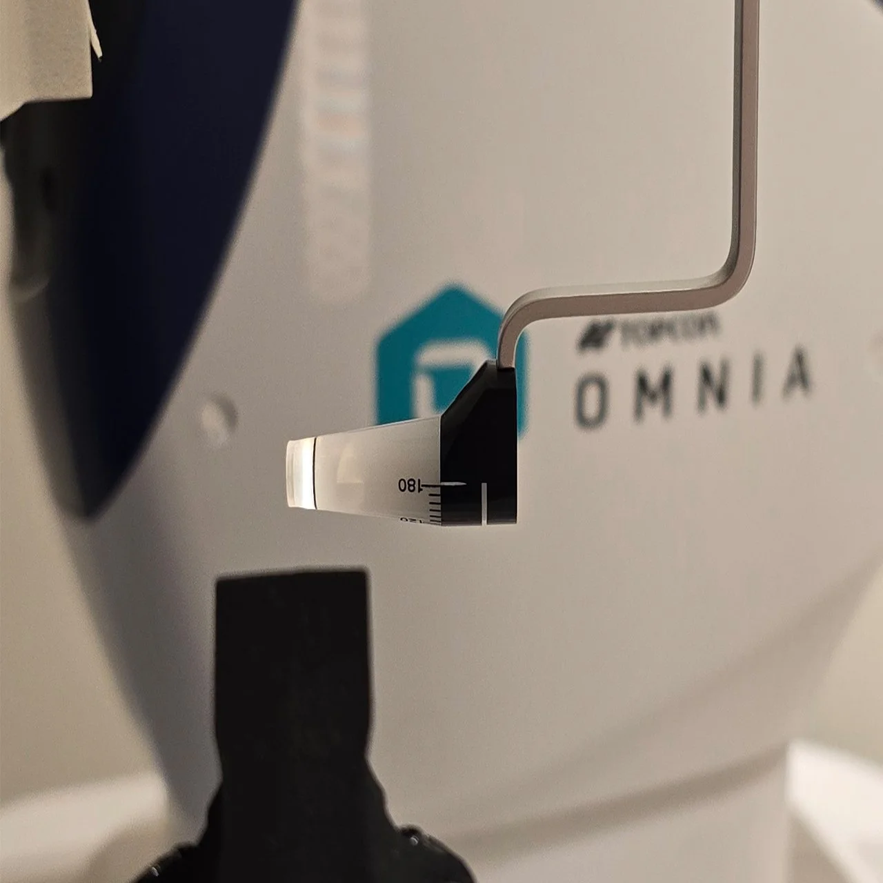
Pomiar ciśnienia wewnątrzgałkowego czyli tonometria to szybkie i bezbolesne badanie, które pomaga we wczesnym wykrywaniu chorób oczu, takich jak jaskra.
W naszym gabinecie wykonujemy pomiar dwiema metodami. Bezkontaktowa tonometria (air-puff) wykorzystuje krótki strumień powietrza i nie wymaga znieczulenia. W razie potrzeby wykonujemy również pomiar tonometrem Goldmanna – uznawanym za najdokładniejszą metodę oceny ciśnienia w oku.
-
Badanie jest wliczone w koszt konsultacji okulistycznej lub może być wykonane osobno wtedy opłata za jego wykonanie wynosi 100 zł
Badanie dna oka
-
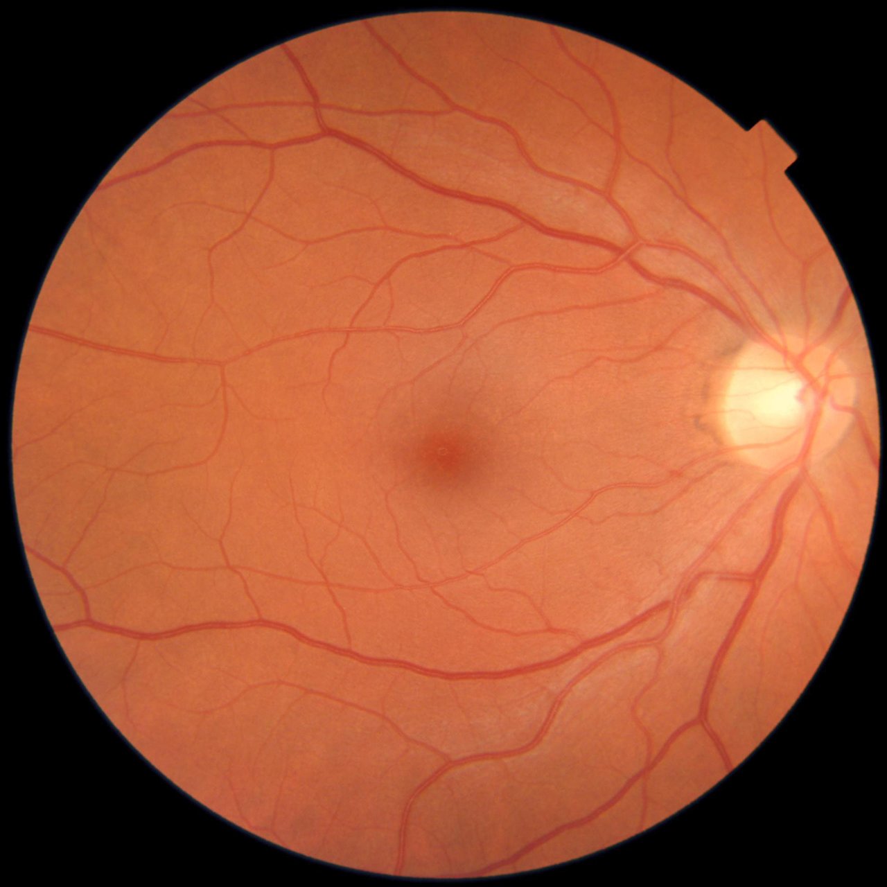
Badanie dna oka pozwala ocenić stan siatkówki, nerwu wzrokowego oraz naczyń krwionośnych znajdujących się wewnątrz oka.
Jest jednym z najważniejszych badań okulistycznych, ponieważ umożliwia wczesne wykrycie wielu chorób oczu, takich jak jaskra, zwyrodnienie plamki żółtej czy zmiany związane z cukrzycą i nadciśnieniem. -
Badanie jest wliczone w koszt konsultacji okulistycznej
Badanie widzenia barwnego
-
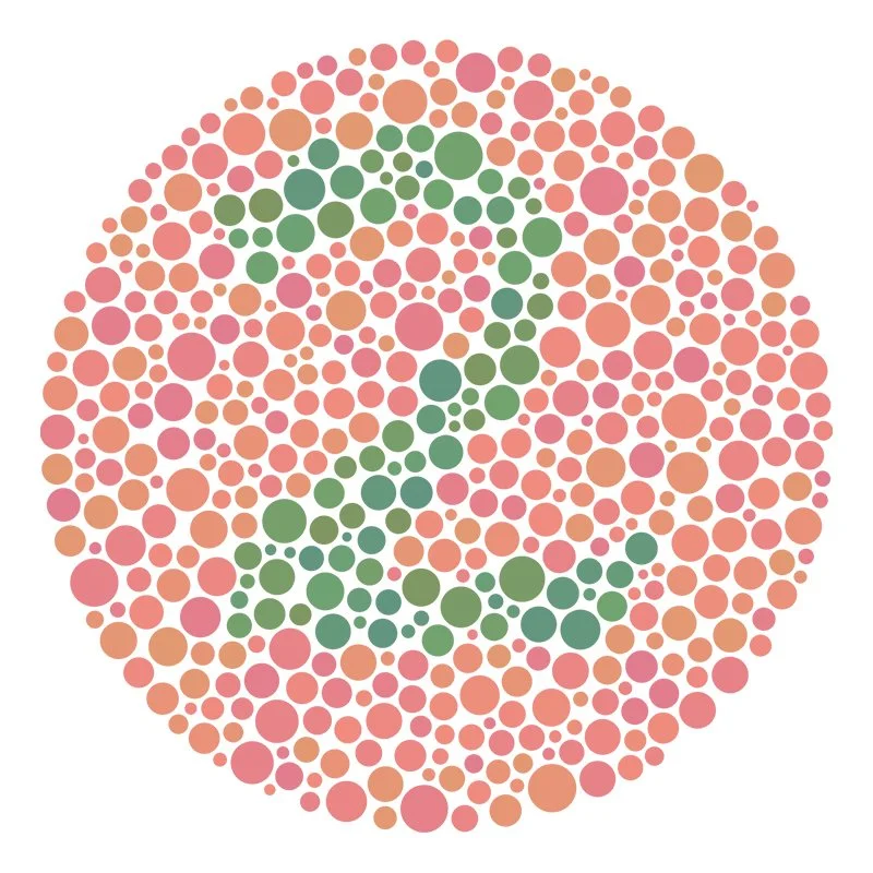
Przesiewowy test służący do wykrywania zaburzeń rozpoznawania barw, szczególnie w zakresie czerwieni i zieleni. Polega na odczytywaniu liczb lub kształtów ukrytych w mozaice kolorowych punktów.
-
Badanie jest wliczone w koszt konsultacji okulistycznej

Badanie widzenia przestrzennego
-
Test Langa to proste, szybkie i bezbolesne badanie, które pozwala ocenić widzenie przestrzenne oraz współpracę obu oczu. Jest wykonywane zarówno u dzieci, jak i dorosłych i pomaga wykryć zaburzenia widzenia obuocznego.
-
Badanie jest wliczone w koszt konsultacji okulistycznej lub może być wykonane osobno wtedy opłata za jego wykonanie wynosi 100 zł
Zdjęcie dna oka
-
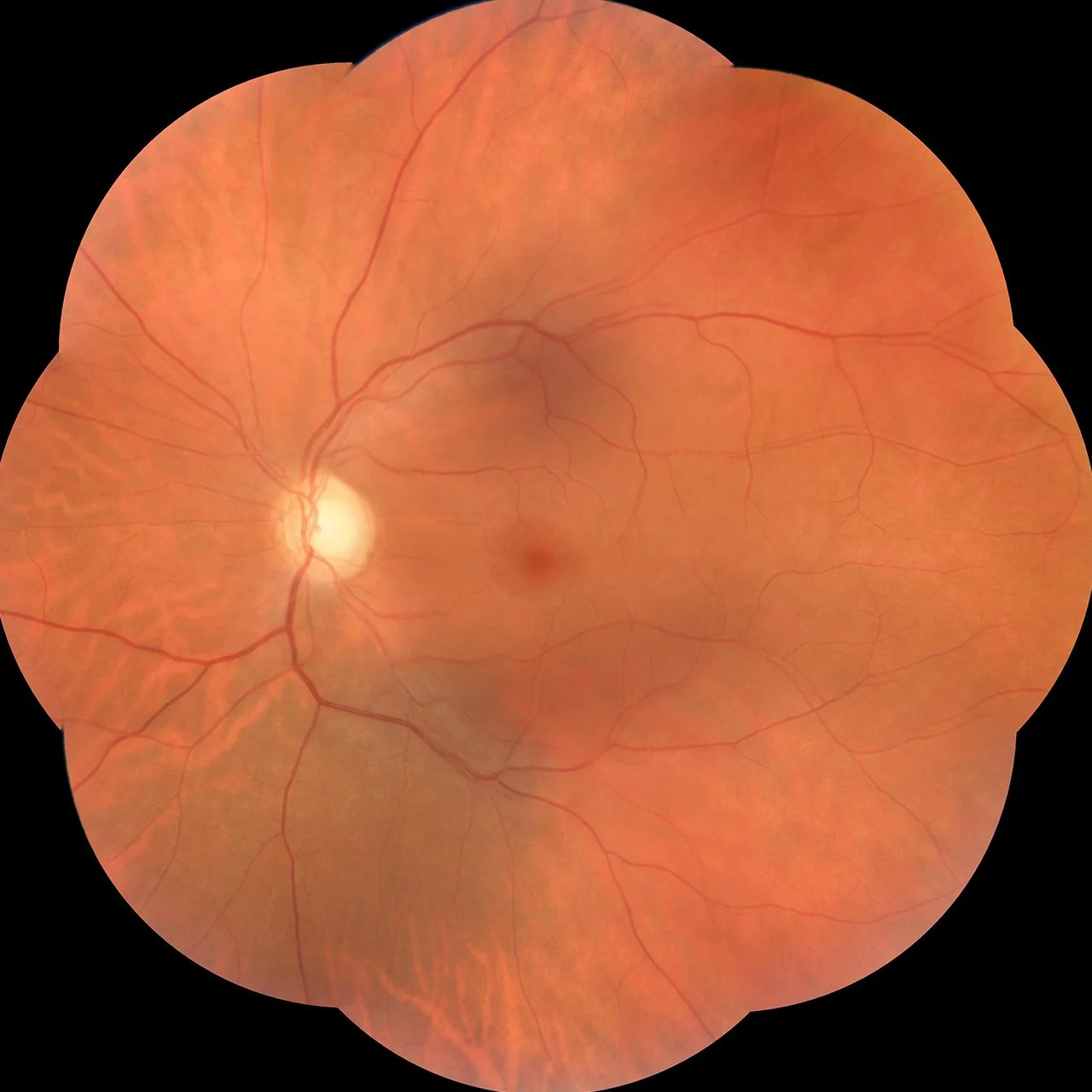
Wykonujemy kolorowe zdjęcia dna oka oraz zdjęcia w świetle bezczerwiennym z wykorzystaniem nowoczesnego aparatu OCT Maestro2. Badanie umożliwia dokładną dokumentację zmian występujących na siatkówce oraz ocenę ich charakteru i rozległości.
Aparat pozwala na wykonanie zdjęć obejmujących pole widzenia 45°. Dodatkowo istnieje możliwość wykonania serii zdjęć w 9 kierunkach, które są automatycznie łączone w panoramiczny obraz dna oka. Dzięki temu uzyskujemy znacznie szerszy zakres obrazowania, co ułatwia ocenę zmian zlokalizowanych również w obwodowych obszarach siatkówki.Badanie dna oka pozwala ocenić stan siatkówki, nerwu wzrokowego oraz naczyń krwionośnych znajdujących się wewnątrz oka.
-
120,00 zł
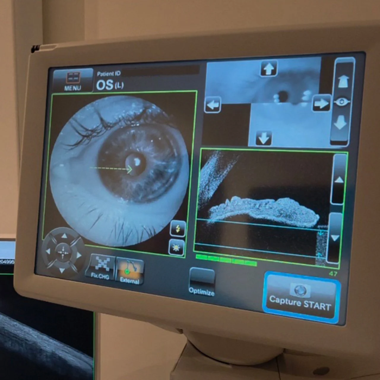
Obrazowanie przedniego odcinka oka
-
Badanie optycznej koherentnej tomografii (OCT) przedniego odcinka oka bez konieczności stosowania dodatkowych soczewek. Dzięki temu badanie jest szybkie, komfortowe i całkowicie nieinwazyjne.
Wykonujemy skany rogówki z pomiarem jej grubości (pachymetria) oraz ocenę i pomiar kąta przesączania. Badania te odgrywają istotną rolę w diagnostyce, monitorowaniu oraz ocenie ryzyka rozwoju jaskry.
-
260,00 zł

Badanie optometryczne
-
Wizyta u optometrysty to kompleksowe badanie wzroku, które pozwala ocenić nie tylko ostrość widzenia, ale również komfort pracy oczu i ich prawidłowe funkcjonowanie.
Badanie rozpoczyna się od szczegółowego wywiadu dotyczącego jakości widzenia, codziennych aktywności oraz ewentualnych dolegliwości. Następnie przeprowadzamy precyzyjny pomiar wady wzroku oraz szereg specjalistycznych testów oceniających m.in. ostrość widzenia, współpracę obu oczu, akomodację i sprawność układu wzrokowego. -
Badanie jest wykonanywane przez optometrystę opłata za jego wykonanie wynosi 180,00 zł
Elektrofizjologia
VEP - Wzrokowe potencjały wywołane
-
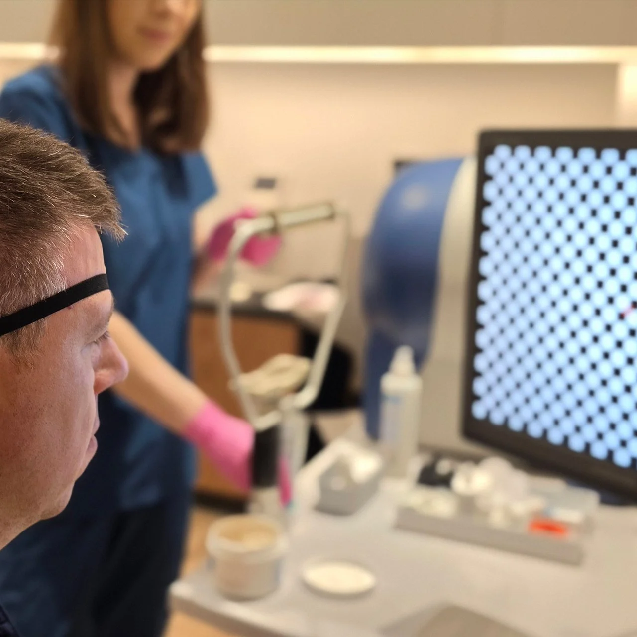
Badania czynnościowe nerwu wzrokowego to wzrokowe potencjały wywołane. Określane są skrótem PVEP (pattern visual evoked potentials), FVEP (flash visual evoked potentials).
U pacjentów z dobrą ostrością wzroku wykonujemy badanie PVEP jako badanie dokładniejsze, u pacjentów z obniżoną ostrością wzroku FVEP, u niektórych pacjentów obydwa badania.
Różnią się one stosowanym bodźcem do stymulacji narządu wzroku. W badaniu pattern jest to biało-czarna szachownica, w badaniu flash- błysk
Główne wskazania:
Choroby nerwu wzrokowego
Zapalenia nerwu wzrokowego w tym również u pacjentów neurologicznych, ze stwardnieniem rozsianym lub jego podejrzeniem
Zanik nerwu wzrokowego częściowy lub całkowity
Obniżenie ostrości wzroku o niewyjaśnionej przyczynie
Ocena funkcji nerwu wzrokowego u dzieci z dużymi wadami wzroku, niedowidzeniem
Psychogenne zaburzenia widzenia
Nieprawidłowy obraz tarczy nerwu wzrokowego.
Przeciwwskazania:
brak współpracy z pacjentem
niektóre postaci padaczki
Badanie wykonuje się bez rozszerzania źrenic, trwa około 15-20 minut.
-
300,00 zł
ERG - Elektroretinografia
-
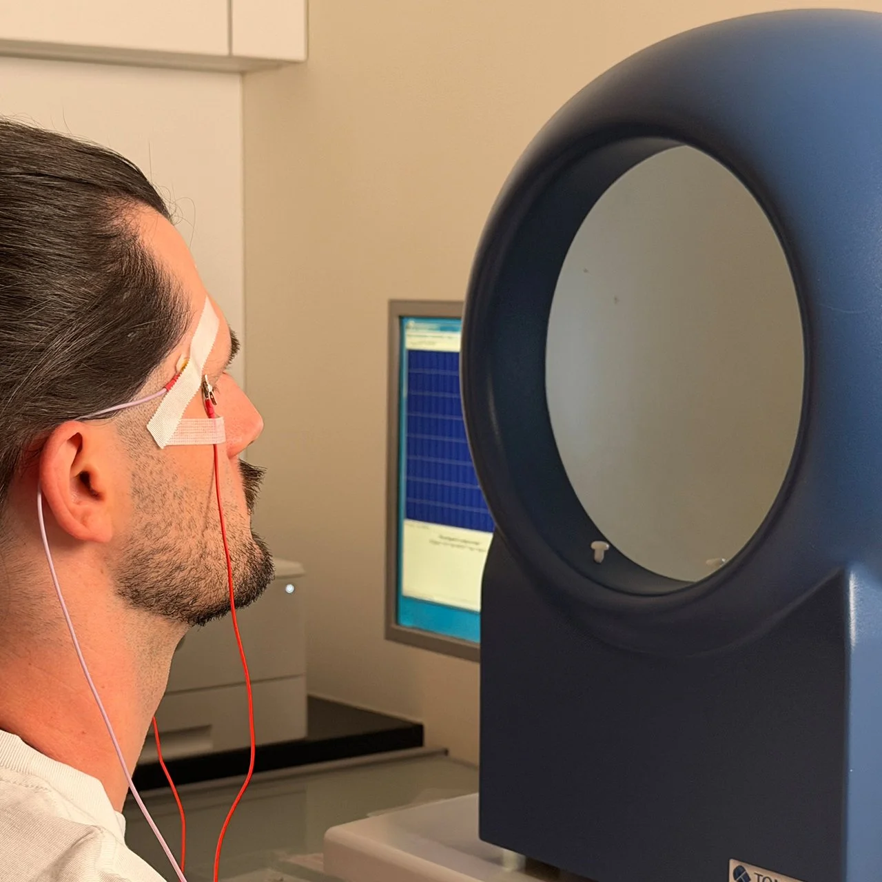
Jest to podstawowe badanie czynnościowe całej siatkówki. Zlecane jest najczęściej przez okulistów u pacjentów z takimi zaburzeniami ze strony narządu wzroku, jak spadek ostrości wzroku o niewyjaśnionej przyczynie, zanurzenia widzenia zmierzchowego, światłowstręt, pewne zaburzenia w polu widzenia. Ponadto elektroretinografia wykonywana jest u pacjentów, u których widzimy pewne charakterystyczne zmiany na dnie oka celem potwierdzenia schorzenia ( np. w zwyrodnieniu barwnikowym siatkówki, chorobie Besta, przebytych stanach zapalnych błony naczyniowej, podejrzeniu ciała obcego wewnątrzgałkowego).
Badanie elektroretinograficzne trwa około 40 minut, jest wykonywane po 20 minutowym okresie adaptacji do ciemności.
Główne wskazania:
choroby plamki i siatkówki wrodzone i nabyte,
genetycznie uwarunkowane – zwyrodnienie barwnikowe siatkówki,
obniżenie ostrości wzroku o niewyjaśnionej przyczynie.
Przeciwwskazania:
brak współpracy z pacjentem,
choroby powierzchni oka.
-
400,00 zł
mfERG - Multifokalne ERG
-
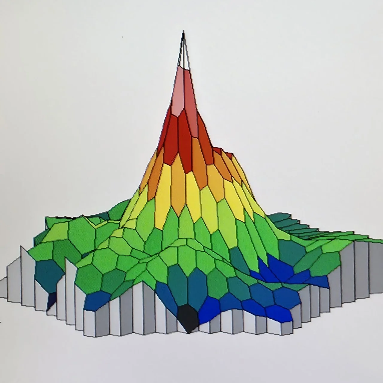
mfERG jest badaniem oceniającym funkcję siatkówki centralnej, 20 stopni od punktu fiksacji.
Badanie to trwa około 45 minut i jest wykonywane po rozszerzeniu źrenic.
Wskazaniem do wykonywania badań elektroretinogramem wieloogniskowym będą:
lokalne uszkodzenia siatkówki,
spadek ostrości wzroku o niewyjaśnionej przyczynie.
Przeciwwskazania:
brak współpracy z pacjentem,
choroby powierzchni oka.
-
380,00 zł

PERG - Elektroretinografia - Pattern
-
Badanie pozwala ocenić pracę komórek nerwowych siatkówki, szczególnie komórek zwojowych, oraz funkcję plamki odpowiedzialnej za ostre widzenie. Badanie pomaga we wczesnym wykrywaniu jaskry oraz jest wykorzystywane w diagnostyce
i monitorowaniu wybranych chorób siatkówki i nerwu wzrokowego. -
350,00 zł
EOG - Elektrookulografia
-
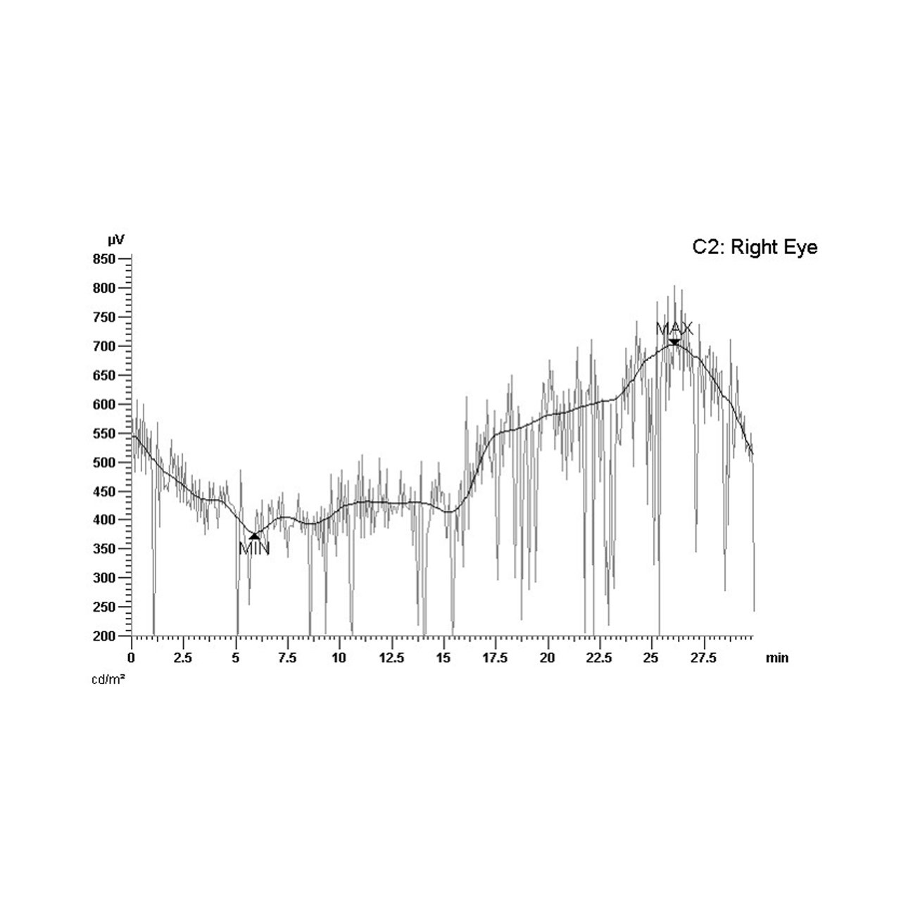
Jest to badanie czynnościowe wybranej warstwy siatkówki - nabłonka barwnikowego. Badanie to najczęściej wykonywane jest w diagnozowaniu chorób u podłoża których leżą nieprawidłowości genu BEST1
Badanie elektrookulograficzne trwa około 40 minut i wykonywane jest po podaniu kropli rozszerzających źrenice
-
420,00 zł
PhNR - Fotopowa odpowiedź negatywna
-
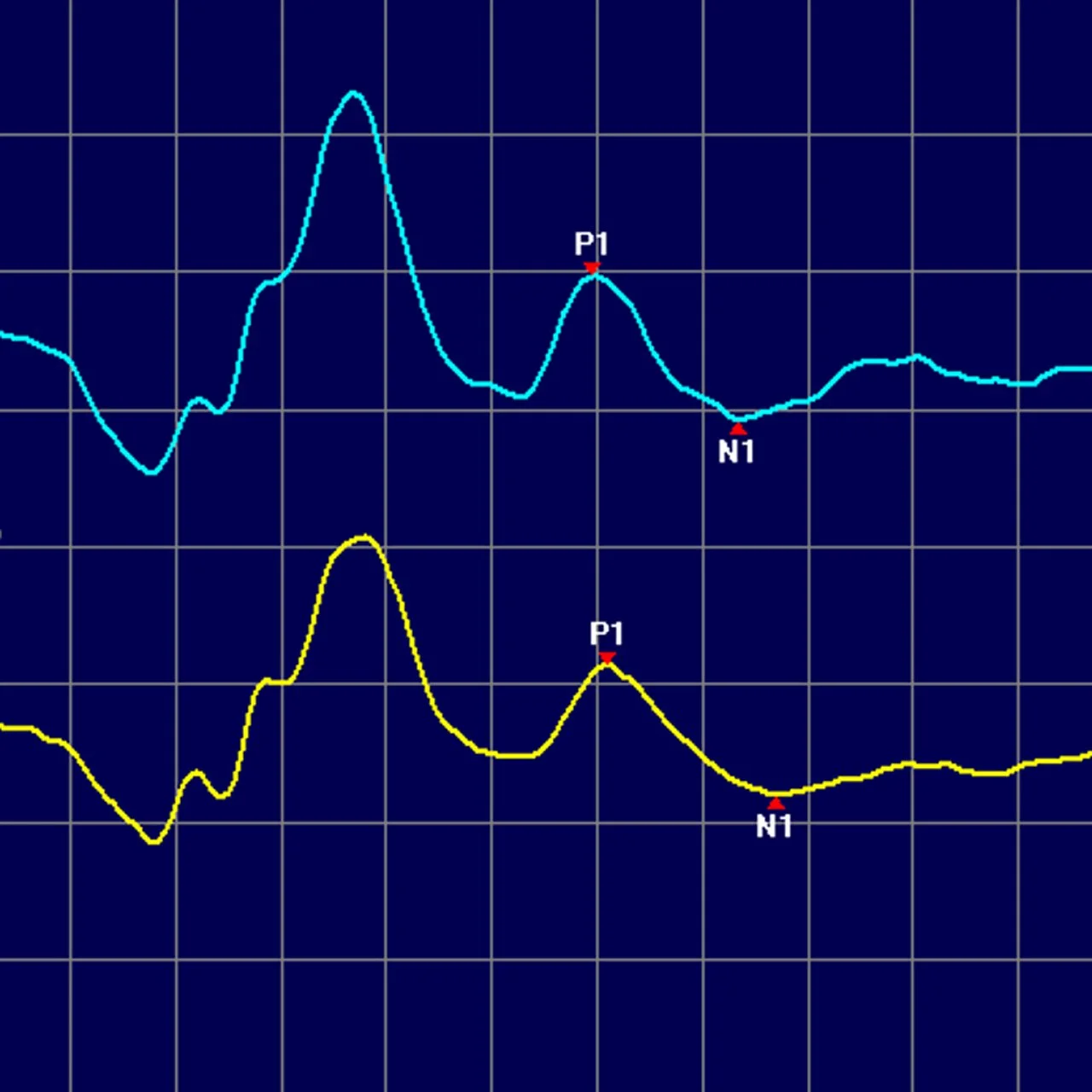
Negatywna odpowiedź fotopowa bada aktywność komórek zwojowych siatkówki. Badanie PhNR stanowi przydatną metodę w procesie diagnostyki jaskry.
-
350,00 zł
Ultrasonografia
USG Doppler tętnic szyjnych i kręgowych
-
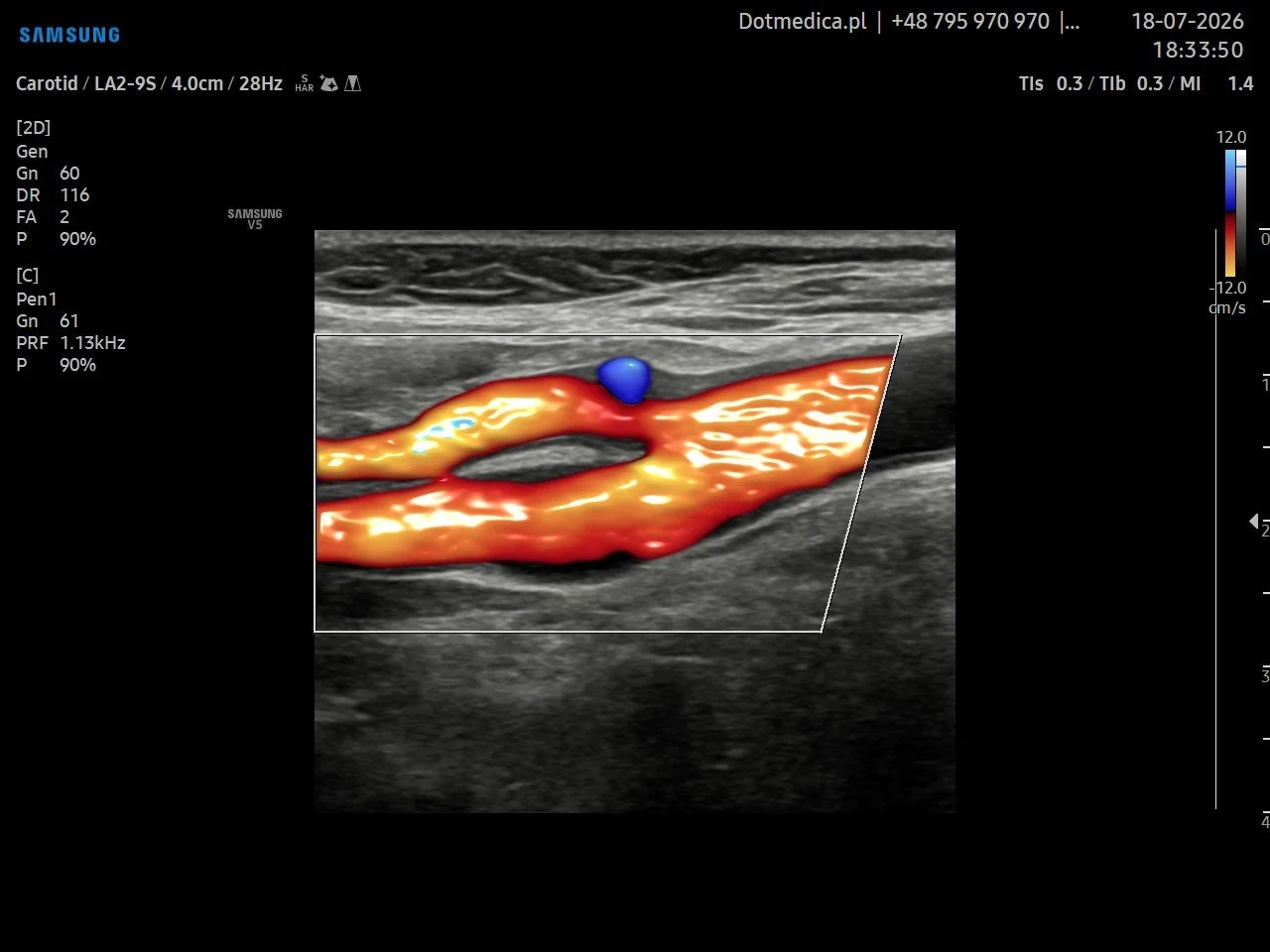
USG Doppler tętnic szyjnych i kręgowych to nieinwazyjne badanie pozwalające ocenić stan naczyń odpowiedzialnych za doprowadzanie krwi do mózgu. Umożliwia analizę przepływu krwi w tętnicach szyjnych oraz kręgowych, a także wykrycie zmian mogących prowadzić do niedokrwienia mózgu.
-
230,00 zł
USG Doppler tętnic i żył kończyn dolnych
-
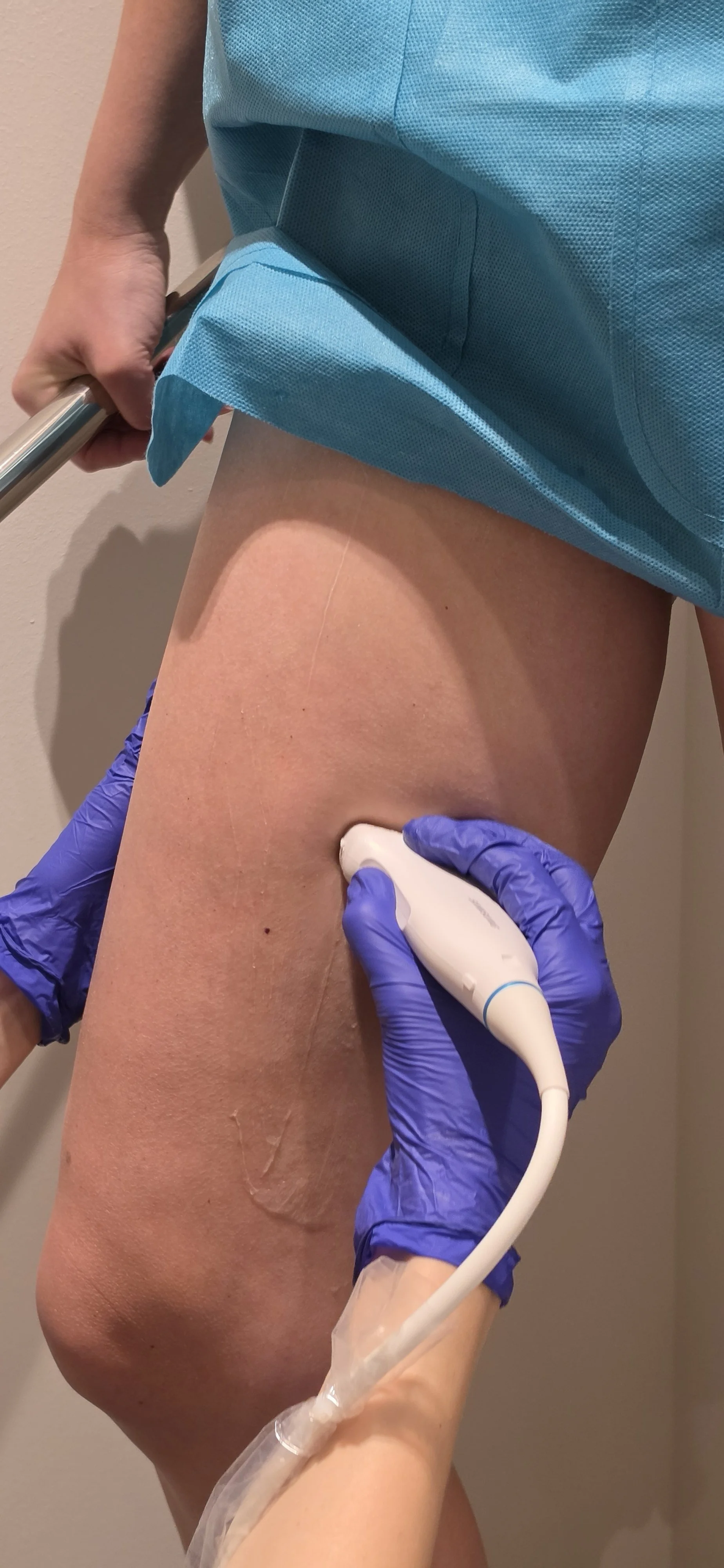
USG Doppler kończyn dolnych to bezbolesne, nieinwazyjne badanie ultrasonograficzne wykorzystujące efekt Dopplera do oceny przepływu krwi w żyłach i tętnicach. Pozwala na wykrycie zakrzepicy, niewydolności żylnej, żylaków oraz miażdżycy.
-
230,00 zł - Doppler tętnic
230,00 zł - Doppler żył350,00 zł - Pakiet żyły + tętnice na jednej wizycie
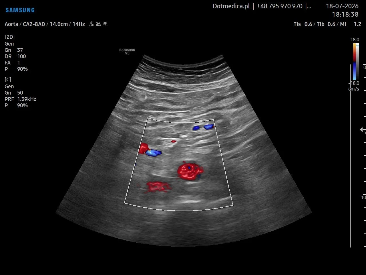
USG doppler aorty brzusznej i tętnic biodrowych
-
USG Doppler aorty brzusznej i tętnic biodrowych to nieinwazyjne i bezpieczne badanie ultrasonograficzne, które umożliwia ocenę aorty – największej tętnicy w organizmie człowieka – oraz przepływu krwi w jej obrębie. Badanie pozwala na dokładną ocenę budowy naczynia, jego średnicy, drożności oraz wykrycie ewentualnych zmian patologicznych.
-
350,00 zł